X-ray tomography in electrocatalysis
2023-10-17 10:00Electrocatalytic background
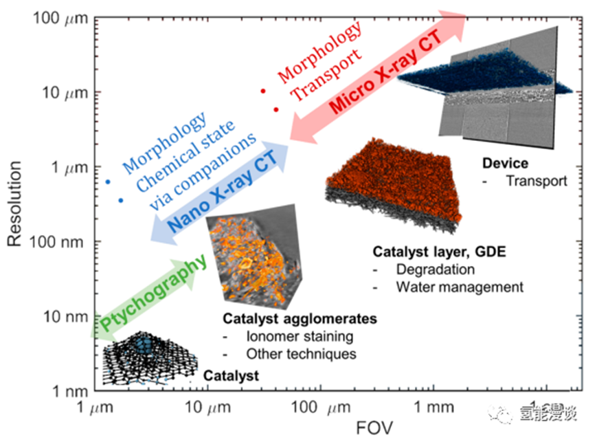
Electrochemical technologies hold great promise for decarbonizing the energy sector and transitioning the economy to net zero. Hydrogen technologies such as fuel cells and electrolyzers are limited by cost and durability. It is necessary to improve the utilization rate, activity and durability of electrocatalysts in order to make them widely used. Similarly, REDOX flow batteries for long-term grid energy storage rely on electrocatalysts for oxidation/reduction reactions, requiring abundant and durable catalysts. These technologies rely on nanoscale electrocatalysts and porous electrodes to increase the surface area and utilization of the catalyst. Overall, Figure 1 summarizes the X-ray CT technique and how it can be applied to study nanoscale and micrometer-scale phenomena related to electrocatalysis in electrochemical devices.

2.Why is X-CT needed for electrocatalysis?
Physicochemical characterization methods such as electron microscopy and X-ray spectroscopy have had a great impact on the development of electrocatalysis. Electron microscopy techniques, such as SEM, TEM, and EDS, can also provide structural and elemental information about the distribution of catalysts within the catalyst layer. In addition, electron-based characterization requires constant maintenance of a high vacuum environment. In contrast, X-ray sources, especially hard X-rays, interact less with gas molecules and require more gentle sample preparation. Therefore, the electrocatalytic community interested in the characterization of electrochemical devices tends to utilize X-ray techniques such as X-ray diffraction, X-ray tomography, and X-ray fluorescence. Micron-scale X-ray tomography is very beneficial for characterizing and understanding electrochemical systems at the micron-scale, and the use of this technique makes an important contribution to understanding the morphology of catalyst layers and their effects on mass transport. As shown in FIG. 3, in the 3D reconstruction results and segmentation results, it can be clearly observed that the platinum anode, the platinum-free cathode, the formation of liquid water in the cathode gap, and the stripping between the platinum catalyst and the expanded film.

