Long time imaging of organisms with micron resolution using X-ray methods
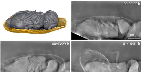
2023-12-29 10:00Researchers have developed an X-ray imaging technique that produces detailed images of organisms at much lower X-ray doses than previously possible.This method is based on phase contrast imaging and depends on the absorption of X-rays by the sample and the wave properties of the X-rays. More precisely, it creates an image from the phase changes that occur as the X-rays pass through the sample.
X-ray imaging can reveal hidden structures and processes inside organisms. However, it also exposes the organism to high doses of harmful radiation, which limits the observation time before damage occurs. To make matters worse, the detection efficiency of commonly used high-resolution detectors decreases as the resolution increases, which means that higher X-ray doses are required to obtain high-resolution images.
To overcome this conundrum, the researchers developed a phase-contrast imaging method that magnifies X-ray images directly. This allows them to use highly efficient large-area detectors while maintaining spatial resolution at the micron level.

The researchers say the approach may also be suitable for biomedical applications, such as gentle tomography examinations of biopsy samples. However, using the Bragg magnifying glass requires a monochromatic, coherent, and collimated beam, which the X-ray synchrotron facility can provide.
